آخر المواضيع المضافة

 النبات
الحيوان
الأحياء المجهرية
علم الأمراض
التقانة الإحيائية
التقنية الحيوية المكروبية
التقنية الحياتية النانوية
علم الأجنة
الأحياء الجزيئي
علم وظائف الأعضاء
الغدد
المضادات الحيوية
النبات
الحيوان
الأحياء المجهرية
علم الأمراض
التقانة الإحيائية
التقنية الحيوية المكروبية
التقنية الحياتية النانوية
علم الأجنة
الأحياء الجزيئي
علم وظائف الأعضاء
الغدد
المضادات الحيوية| Capillary Network-Lacrimal Gland |


|
|
Read More
Date: 31-7-2016
Date: 9-1-2017
Date:
|
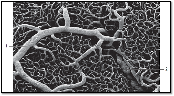
Capillary Network-Lacrimal Gland
The capillary bed of an organ can be made visible using one of several injection techniques, and its three-dimensional structure can then be examined by scanning electron microscopy. In this instance, the arteries that go to the head of a cat were filled with a resin, which was allowed to harden at a defined temperature range. Organic materials were than removed using acid or alkaline solutions (maceration ). This procedure creates a corrosion preparation, which leaves the geometry of the capillaries intact. This figure shows the dense capillary network in the lacrimal gland of a cat. Note the branching of the larger arteries 1 . Veins 2 are visible in the bottom left of the figure. Differences in form and orientation of the endothelial cell nuclei, among other attributes, indicate whether a vessel is an artery or vein.
1 Arteries
2 Veins
Scanning electron microscopy; magnification: × 85
References
Kuehnel, W.(2003). Color Atlas of Cytology, Histology, and Microscopic Anatomy. 4th edition . Institute of Anatomy Universitätzu Luebeck Luebeck, Germany . Thieme Stuttgart · New York .

|
|
|
|
للعاملين في الليل.. حيلة صحية تجنبكم خطر هذا النوع من العمل
|
|
|
|
|
|
|
"ناسا" تحتفي برائد الفضاء السوفياتي يوري غاغارين
|
|
|
|
|
|
|
ملاكات العتبة العباسية المقدسة تُنهي أعمال غسل حرم مرقد أبي الفضل العباس (عليه السلام) وفرشه
|
|
|