

 النبات
 الحيوان
 الأحياء المجهرية
 علم الأمراض
 التقانة الإحيائية
 التقنية الحيوية المكروبية
 التقنية الحياتية النانوية
 علم الأجنة
 الأحياء الجزيئي
 علم وظائف الأعضاء
 الغدد
الغدد
 المضادات الحيوية
| Granular (Rough) Endoplasmic Reticulum (rER)—Ergastoplasm—Nissl Bodies |
|
|
|
Read More
Date: 23-1-2017
Date: 18-1-2017
Date: 1-8-2016
|
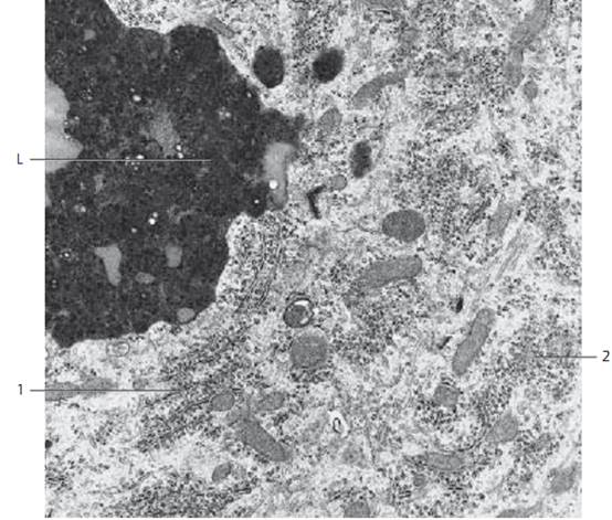
Granular (Rough) Endoplasmic Reticulum (rER)—Ergastoplasm—Nissl Bodies
The bluish-violet bodies or patches ( Nissl bodies, Nissl substance) are Ultrastructurally identical to the highly developed ergastoplasm ( rER) 1 , which consists of anastomosing stacked double membranes with bound ribosomes, rER-derive d cisternae as well as regions with many free ribosomes 2 .
Ribosomes are the smallest cell organelles. With a diameter of about 25 nm, they are clearly visible as ribonucleoprotein particles in transmission electron microscopy. Ribosomes are involve d in the biosynthesis of proteins, includ-ing secretory, lysosomal and membrane-bound proteins. They consist of a large and a small subunit . Free ribosomes are present in large numbers in the cytoplasmic matrix, either as single ribosomes or in smaller groups (polysomes ). It depends on the type of neuron whether groups of free polysomes or rER-bound ribosomes pre dominate. While G. E. Palade described ribosomes as particles with high affinity to contrast dyes as early as 1955, the name “ribosome” was introduced later by R. B. Roberts in 1958. This picture shows part of a Purkinje cell from the cerebellum. In the perikar yon are crista-type mitochondria and neurotubules.
1 rER
2 Ribosomes
L Lipofuscin granule

References
Kuehnel, W.(2003). Color Atlas of Cytology, Histology, and Microscopic Anatomy. 4th edition . Institute of Anatomy Universitätzu Luebeck Luebeck, Germany . Thieme Stuttgar t · New York .


|
|
|
|
للعاملين في الليل.. حيلة صحية تجنبكم خطر هذا النوع من العمل
|
|
|
|
|
|
|
"ناسا" تحتفي برائد الفضاء السوفياتي يوري غاغارين
|
|
|
|
|
|
|
المجمع العلمي يقيم ورشة تطويرية ودورة قرآنية في النجف والديوانية
|
|
|