

 النبات
 الحيوان
 الأحياء المجهرية
 علم الأمراض
 التقانة الإحيائية
 التقنية الحيوية المكروبية
 التقنية الحياتية النانوية
 علم الأجنة
 الأحياء الجزيئي
 علم وظائف الأعضاء
 الغدد
الغدد
 المضادات الحيوية
|
Read More
Date: 27-7-2021
Date: 16-7-2021
Date: 18-7-2021
|
Joints and Ligaments of the upper limb
Joints of the upper limb include all joints of the shoulder complex, elbow (humeroradial and humeroulnar), forearm (proximal and distal radioulnar), and hand (wrist, intercarpal, CMC, MCP, and IP). A series of ligaments and capsular structures support these upper limb joints and permit a wide range of movement.
A. Shoulder complex
Shoulder complex joints include the glenohumeral, acromioclavicular, and sternoclavicular joints (Figs. 1 and 2).

Figure 1 : Glenohumeral joint.

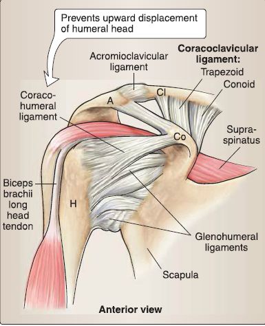
Figure 2 : Capsular and ligamentous support of shoulder complex.
1. Glenohumeral: This is a synovial ball-and-socket joint between the head of the humerus and shallow glenoid fossa. It is supported by a joint capsule, glenohumeral and coracohumeral ligaments, and rotator cuff muscles. The joint is deepened by fibrocartilaginous glenoid labrum.
a. Movement: This joint permits flexion, extension, abduction, adduction, medial rotation, lateral rotation, and circumduction. Its wide range of motion makes it vulnerable to injury.
b. Blood supply: This joint is supplied by anterior and posterior circumflex humeral arteries and suprascapular, axillary, and lateral pectoral nerves.
c. Associated bursae: These include the subacromial/subdeltoid and subscapular bursae.
2. Acromioclavicular: This is a synovial plane joint between the acromion and the acromial end of the clavicle. It is supported by a joint capsule, directly by the acromioclavicular ligament and indirectly by the coracoclavicular ligament (conoid and
trapezoid portions).
a. Movement: This joint permits rotation of the acromion on the clavicle during scapular movement.
b. Blood supply: This joint is supplied by branches of the suprascapular and thoracoacromial arteries and supraclavicular, lateral pectoral, and axillary nerves.
3. Sternoclavicular: This is a synovial saddle joint (functionally ball and socket) between the manubrium and the sternal end of the clavicle. It is supported by a joint capsule and anterior and posterior sternoclavicular, interclavicular, and costoclavicular ligaments. It has an articular disc.
a. Movement: This joint permits clavicular elevation, depression, and anterior/posterior movement during humeral movement.
b. Blood supply: This joint is supplied by branches of the internal thoracic and suprascapular arteries and medial supraclavicular and subclavius nerves.
B. Elbow
The elbow is a synovial hinge-shaped joint, supported by a fibrous capsule. It allows for flexion and extension movements and is supplied by anastomoses around joint (from brachia!, profunda brachii, radial, and ulnar arteries) and median, radial, and ulnar nerves .
1. Humeroradial: This joint articulates between the capitulum and radial head and is supported by radial collateral and anular ligaments (also supports proximal radioulnar joint by maintaining position of radial head in radial notch of ulna).
2. Humeroulnar: This joint articulates between the trochlea and trochlear notch and is supported by the ulnar collateral ligament (anterior, posterior, and oblique bands).
C. Forearm
Synovial pivot-type joints allow for pronation and supination of the forearm. They are supported by a fibrous capsule and ligaments.
1. Proximal radioulnar: This joint articulates between the radial head and radial notch of the ulna and allows for rotation of the head in the notch. It is supported by the anular ligament and supplied by elbow anastomoses and musculocutaneous, median, and radial nerves.
2. Distal radioulnar: This joint articulates between the ulnar head and ulnar notch of the radius with an articular disc. The ulna is relatively fixed, allowing the radius to move during pronation/supination. It is supported by weak anterior and posterior ligaments and the interosseous membrane and supplied by anterior and posterior interosseous arteries and nerves.
D. Wrist and hand
The wrist and hand are made up a collection of small synovial joints that permit fine movement at the wrist and fingers, including flexion, extension, abduction, adduction, and opposition (first and fifth digits only). Collectively, these joints are supplied by the carpal arterial arches and associated digital branches and radial, median, and ulnar nerves.
1. Wrist: The radiocarpal joint articulates between the radius and proximal row of carpal bones (minus pisiform). The ulna does not contribute to this joint, which is supported by radial and ulnar collateral ligaments of the wrist and radiocarpal ligaments (palmar and dorsal).
2. Hand: The hand is a collection of intercarpal, CMC, MCP, and IP joints that are supported by fibrous capsules and ligaments (named for joint). The Pisohamate ligament creates an osseofibrous tunnel (Guyon's canal) through which the deep branch of the ulnar nerve travels, a potential site for nerve compression.


|
|
|
|
"عادة ليلية" قد تكون المفتاح للوقاية من الخرف
|
|
|
|
|
|
|
ممتص الصدمات: طريقة عمله وأهميته وأبرز علامات تلفه
|
|
|
|
|
|
|
ندوات وأنشطة قرآنية مختلفة يقيمها المجمَع العلمي في محافظتي النجف وكربلاء
|
|
|