Interpretation of spectra
The number of fundamental vibrational modes of a molecule is equal to the number of degrees of vibrational freedom. For a nonlinear molecule of n atoms, there are 3n 6 degrees of vibrational freedom. Hence, there are 3n 6 fundamental vibrational modes. The number of degrees of freedom is reduced by 6 for a nonlinear molecule because:
• three coordinates are required to locate the molecule in space
• an additional three coordinates are required to describe the orientation of the molecule in the position based defined by the three coordinates that locate the molecule in space.
For a linear molecule, 3n 5 fundamental vibrational modes are possible, since only two degrees of rotational freedom exist. Thus, in a total vibrational analysis of a molecule by complementary IR and Raman techniques, 3n 6 or 3n 5 vibrational frequencies should be observed. In complex molecules (such as pharmaceuticals), it may not be possible to observe every vibration because of overlap and relative intensity differences. Regarding spectral interpretation, two of the best textbook sources are Introduction to Infrared and Raman Spectroscopy (Colthup et al. 1990) and The Handbook of Infrared and Raman Characteristic Frequencies of Organic Molecules (Lin-Vien et al. 1991). Both of these texts contain chapters dedicated to functional groups (e.g. methyl and methylene groups) and chemical compound types (e.g. ethers, alcohols and phenols). By examining peak location and intensity in Raman spectra, functional groups (and chemical compound type) can be determined. Discussed below are some guidelines on interpretation of a Raman spectrum, but clearly these serve only as general background information. Any complete spectral interpretations should be made with the guidance of one of the reference texts.
Raman spectroscopy provides information about the molecular bonding of a molecule. Certain functional groups give rise to funda mental vibrational modes. For example, the C=C stretch inethylenes occurs near 1650 cm1. The C-N stretch (amide III peak) for primary amides occurs between 1430 and 1390 cm1. A C-S stretch usually appears strongly in the 735 to 590 cm-1 region. Many other functional groups give rise to Raman peaks in specific spectral regions. IR bands also occur in the same spectral regions, but because of selection rules, the band intensities differ, often dramatically. As noted above, in the most basic terms, a vibrational mode is Raman active when there is a change in polarisability during the vibration.
Conversely, a vibrational mode is IR active when there is a change in the molecular dipole moment during the vibration. Hence, vibra tional modes that give rise to strong Raman peaks often give weak IR bands and vice versa. It is this characteristic that provides the description ‘complementary’ for the use of IR and Raman spectroscopy together to examine the molecular bonds in a compound. Some of the strongest Raman peaks come from functional groups such as C=C, N=N, S-S, C-H, S-H, C=N, C=S and C-S, which have low polarity and high polarisability. These functional groups tend to occur in rather constant frequency ranges, although some shifts can occur from the influence of other substituents in the molecule. Vibrational modes can be separated into two classes: those in which the molecular bond is stretching and those in which it is bending. It takes a specific amount of energy for these actions to occur. The amount required is dependent on the atoms involved and the strength of the bond. The nature of Raman excitation of a molecule is discussed in the theory section at the beginning of this chapter. The location of the vibrational mode (its peak wave length position in the spectrum) is related to the frequency of the excitation source (the laser) and the frequency of the scattered light. The equation that defines this relationship is
hV=h̷v+∆Evib
where h is Plank’s constant, v is the excitation frequency, t is the scattered light frequency and ∆Evib is the vibrational energy. The vibrational energy is related directly to the strength of the bond and the amount of energy required to make that bond stretch or bend. These actions are elaborated below.
A stretching vibration is the motion a molecular bond undergoes when the two atoms involved in the bond move apart and then contract. The stretch can be a simple contraction–expansion between two atoms, such as the C=C stretch of ethylene. This type of motion occurs as a peak in the Raman spectrum at approximately 1650 cm-1. When three atoms are involved, two types of stretches are possible, symmetric and antisymmetric. Using a methylene group as an example, a symmetric CH2 stretch occurs when the two hydrogen atoms move apart from the carbon atom at the same time. This type of stretch appears near 2853 cm1. The antisymmetric CH2 stretch occurs when one hydrogen atom moves away from the carbon atom while the other hydrogen atom moves closer to the carbon atom. This vibrational mode appears near 2926 cm-1. The symmetric stretches are typically more intense in Raman spectra (greater change in polarisability), whereas the antisymmetric stretches are more intense in IR spectra. When many atoms are involved in the stretching, such as with aromatic rings, the types of stretches become more complicated. For example, a benzene ring monosubstituted with a halogen has 30 vibrational modes. Of these modes, some are strictly stretches, some are bends and some are combinations of both. Perhaps the simplest stretching mode for benzene rings is the ring breathing mode, in which the 2-, 4- and 6 carbons move outwards. This mode occurs as a very strong Raman peak near 1000 cm-1 for mono-, meta- and 1,3,5-trisubstituted benzenes. Clearly, many types of stretches can occur.
In a bending vibration, the molecular bond bends instead of stretches. Many types of bends can occur: antisymmetric, symmetric, torsion, scissor, wag, twist and rock. Using a CH3 group as an example, antisymmetric, symmetric, rock and torsion bends are possible (Fig. 1). In the anti-symmetric bend, two of the C-H bonds bend towards each other in a pinching motion, while the third bends outwards and away from the pyramid. This mode occurs between 1470 and 1430 cm-1. In the symmetric bend, the three CH bonds all bend inwards, similar to a grasping action. This mode occurs from 1395 to 1365 cm1. In the CH3 rock, the three CH bonds all bend in one direction, in a sweeping mode. Finally, the CH3 torsion involves the three CH bonds all bending in a clockwise direction in a twisting motion. The spectral ranges for these last two vibrational modes are highly variable.
The CH2 group can bend in slightly different ways . A CH2 scissor bend occurs when the two hydrogen atoms move towards each other in a scissoring motion. For a CH2 wag, the two hydrogen atoms are bent towards the carbon atom. If the two hydrogen atoms alternately twist around one another, it is a CH2 twist. Finally, if the two hydrogen atoms move back and forth, in line with the carbon atom, it is a CH2 rock. As seen with the CH3 modes, all of these different types of motions appear in different portions of the spectrum because they each require a different and unique amount of energy to occur. The scissor mode appears near 1465 cm1. The rocking, wagging and twisting modes are more complicated, falling in the range 1422–719 cm-1. Depending on the substituents in the molecule, each of these modes can be narrowed down to tighter ranges. It is this dependence of certainmodeson substituents that gives rise to correlation tables, discussed next. Although the general location for a certain type of mode, for example a C=C stretch, can be listed, the exact location of the peak varies slightly with the type of molecular substitution present. For example, a vinyl C=C stretch (monoalkyl) occurs from 1650 to 1638 cm1. A vinylidine C2C stretch (1,1-dialkyl) occurs from 1660 to 1640 cm1.Acis-dialkyl-substituted C2C stretch occurs from 1662 to 1631 cm-1, whereas a trans-dialkyl-substituted C=C stretch occurs from 1676 to 1665 cm1. Finally, tri alkyl- and tetraalkyl-substituted C=C stretches appear from 1680 to 1665 cm1. Some modes are very sensitive to substituents, whereas others appear relatively consistently near a certain wavenumber.
Over the years, many types of compounds have been studied in great detail. When a spec trum of a single compound is interpreted such that every peak or band is assigned to a type of motion, we have a vibrational assignment. Through the examination of many vibrational assignments, it has been possible to draw correlations between types of vibrational modes and types of substituents. The culmination of this work is detailed in correlation tables that an investigator can use to aid in spectral interpretation (Dollish et al. 1974; Colthup et al. 1990; Lin-Vien et al. 1991).
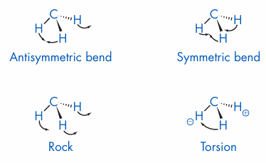
Figure 1 Bending vibrational motions associated with CH3 groups (+ represents movement above the plane, - represents movement below the plane).
The most efficient way to use correlation tables is to look for certain peaks that will quickly narrow down the type of compound. For example, if peaks are found at 3000 cm1 or slightly higher, the compound is aromatic or olefinic. However, if the CH2 stretches appear below 3000 cm1, the compound is aliphatic. Once that determination is made, the correlation tables can be consulted to look for other confirmatory modes, such as CH wags for aromatic compounds. This type of process is continued until a compound class can either be verified or discounted. For complex molecules, it may not be possible to determine the structure from the Raman spec trumalone, or even if the IR spectrum is available as well. Other techniques such as NMR and mass spectrometry provide important information to aid the elucidation of structure.








