آخر المواضيع المضافة

 النبات
الحيوان
الأحياء المجهرية
علم الأمراض
التقانة الإحيائية
التقنية الحيوية المكروبية
التقنية الحياتية النانوية
علم الأجنة
الأحياء الجزيئي
علم وظائف الأعضاء
الغدد
المضادات الحيوية
النبات
الحيوان
الأحياء المجهرية
علم الأمراض
التقانة الإحيائية
التقنية الحيوية المكروبية
التقنية الحياتية النانوية
علم الأجنة
الأحياء الجزيئي
علم وظائف الأعضاء
الغدد
المضادات الحيوية| Single-Layered Pseudostratified Columnar Epithelium-Duodenum |


|
|
Read More
Date: 9-1-2017
Date: 17-1-2017
Date: 25-7-2016
|
Single-Layered Pseudostratified Columnar Epithelium-Duodenum
In single-layered Pseudostratified epithelium, the longitudinal axis of the cells is always oriented vertical to the tissue surface. The cells appear polygonal in cross-sections. A row of oval nuclei mostly occupy the basal part of the cell, while most of the cell organelles are locate d in the supranuclear cell region. The free surfaces of the epithelial (enterocytes ), cells in this figure have a clearly visible striate d border 1 which consists of microvilli. Sporadically, goblet cells 2 occur intersperse d with the epithelial cells of the tissue. The cells of the lamina propria mucosae 3 form a connective tissue layer underneath the epithelium, which also contains smooth muscle cells 4 , apart from blood and lymph vessels, nerve fibers and myofibroblasts. A thin basal membrane (stained blue in this section) separates the epithelium from the lamina propria mucosae 3 .
1 Brush border
2 Goblet cells
3 Lamina propria mucosae
4 Smooth muscle cells
5 Terminal web
Stain: azan; magnification: × 400

Vertical section through the lamina epithelial is mucosae of the duodenum to describe single-layered Pseudostratified columnar epithelium . The slender Pseudostratified epithelial cells are evenly covered with a brush border 1. The oval nuclei are locate d in the basal third of the cell. All enterocytes contain a large number of mitochondria, both in the perinuclear (basal) space and in the apical third of the cell 2 .
Microvilli, here seen as brush border, enlarge the duodenal surface and allow considerably more contact with the intestinal content. Microvilli-associated enzymes are important for the resorption of nutrients and digestion. The lamina propria mucosae 3 is visible in the lower part of the figure.
1 Brush border (microvilli)
2 Mitochondria
3 Lamina propria mucosae
4 Intestinal lumen
Electron microscopy; magnification: × 2000

References
Kuehnel, W.(2003). Color Atlas of Cytology, Histology, and Microscopic Anatomy. 4th edition . Institute of Anatomy Universitätzu Luebeck Luebeck, Germany . Thieme Stuttgar t · New York .

|
|
|
|
دخلت غرفة فنسيت ماذا تريد من داخلها.. خبير يفسر الحالة
|
|
|
|
|
|
|
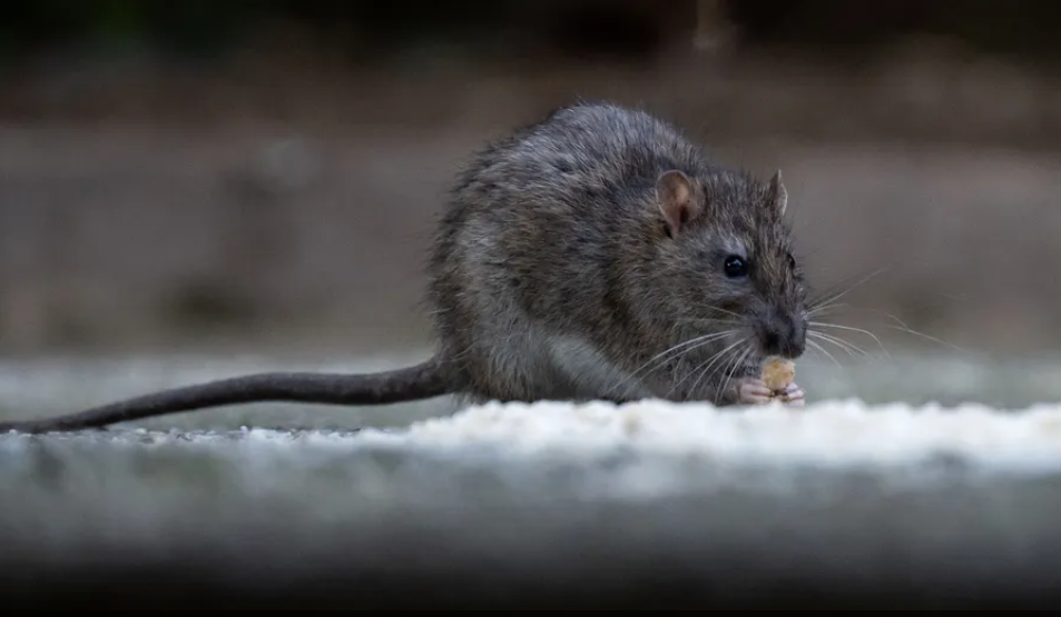
دراسة: الفئران تستخدم "قبلة الحياة" لإنعاش أقرانها
|
|
|
|
|
|
|
أصواتٌ قرآنية واعدة .. أكثر من 80 برعماً يشارك في المحفل القرآني الرمضاني بالصحن الحيدري الشريف
|
|
|