

 النبات
 الحيوان
 الأحياء المجهرية
 علم الأمراض
 التقانة الإحيائية
 التقنية الحيوية المكروبية
 التقنية الحياتية النانوية
 علم الأجنة
 الأحياء الجزيئي
 علم وظائف الأعضاء
 الغدد
الغدد
 المضادات الحيوية
|
Read More
Date: 4-11-2015
Date: 4-11-2015
Date: 4-11-2015
|
Pigeon
Sub phylum - Vertebrata
Class - Aves
Order - Columbiformes
Type - Columba livia
Birds are easily recognized group of vertebrates. In birds every part of the body is modified to suit their aerial mode of life. Birds possess feathers, beak and feet modified in relation to their aerial life.
The Pigeons are flying birds (carinate). They are known both as wild and domesticated forms. The Pigeons are seen both in tropical and temperate zones. About 10 species of Pigeons are found in India. The pigeons fly in flocks and roost together. The domestic pigeons have many varieties, namely panter, fantail and tumblers. They differ in size, colorations and feather arrangement. All of them are, however, descendants of the rock pigeon-columba livia
External features:

The Body is spindle shaped. Their size varies from 20-25 cm. They are covered by coloured feathers leaving beak and a small portion of the hindlimbs.
The body is divisible into head, neck, trunk and a small, conical tail. The head is round and drawn out anteriorly into a strong, hard, pointed beak. The mouth is a terminal wide gape, guarded by elongated upper and lower beaks. The beaks are covered with a horny sheath or rhampotheca. A swollen area of soft skin, the cere, surrounds the nostril. It is present on each side of the upper beak. The eyes are large and guarded by upper and lower eyelids and a transparent nictitating membrane. A pair of ear openings are situated at a short distance behind the eyes. Each opening leads into a short external auditory meatus, ending in the tympanic membrane forming the ear drum.
The neck is long and mobile. It helps in the movement of the head in various directions. The trunk is compact, heavy and bears a pair of wings and a pair of legs. The cloacal aperture is at its hind end on the lower surface. Projecting behind the cloacal aperture is the tail. Above the tail is a knob on which opens an oil gland or preen gland or uropygeal gland. It secretes a fluid used for preening the feathers.
The wings:

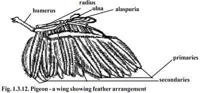
The forelimbs as modified wings are located in the anterior region of the trunk. The limbs are of the pentadactyl type. The wing has three typical divisions as - the upper arm, forearm and hand. The hand has three imperfectly marked digits. While the pigeon is at rest the three divisions of the wing are bent upon one another in the form of the letter ‘Z’. During flight the wings are straightened and extended. A fold of skin the alar membrane or prepatagium, stretches between the upper and forearm along the anterior border of the limb. A smaller fold known as postpatagium is present between the trunk and upperarm.

While the pigeon is not flying the whole weight of the body has to be supported by the hind limbs, In order to balance the heavy trunk the hindlimbs are attached for forwards. Each hindlimb or leg has three typical divisions, the thigh, shank and foot. The thigh without being free is enclosed within the boundaries of the trunk. Each hindlimb has four digits. The first toe is directed backward. The feet are naked and covered with horny epidermal scales. Each digit is provided with a horny claw. The tail is small and concealed by the feathers of the trunk. It bears the tail feathers or rectrices.
Exoskeleton
The feathers are integumentary structures. They are characteristic of birds. The feathers are derived from the epidermis. They are seasonally or periodically cast off and the new one being developed from the old papillae. They are arranged on the skin in definite tracts, called feather tracts or pterylae. The interspaces without feathers are known as apteria or featherless tracts.
There are three types of feathers in pigeon. They are the large quill feathers found on wings and tail for flight, the contour feathers, forming a covering for the body and the filoplumes, lying between the contour feathers.

Quill feather:
Each quill feather has a central stem or scapus. It is divided into lower hollow part called the quill or calamus and a solid upper part termed rachis. The quill has at its lower ended an opening called inferior umbilicus, through which vascular processes or papilla of the dermis project into the growing feather. Another opening the Superior umbilicus occurs at the junction of quill and the rachis on the inner face of feather. Close to this opening, there is a small tuft of soft feathers called aftershaft. Attached to the rachis are small filaments or barbs. The rachis with the barbs constitute the vane or vexillum. Each barb is provided with barbules and hooklets. The barbs remain attached with one another to form a continuous blade for striking the air in flight.
There are twenty three quill feathers or remiges in each wing. Eleven of these known as primaries. Are attached to the hand. The remaining twelve fixed on the forearm are called secondaries. Attached to the thumb is a small tuft of feathers known as ala spuria or bastard wing. The tail bears twelve tail feathers or rectrices which are arranged in the form of a fan.
The contour feathers are soft and the barbs are plume like with no interlocking mechanisms. These help to keep the body warm and lock air pockets. The filoplumes have delictae hair like long axis and a few barbs devoid of barbules. Down feathers have small axis and a few barbs devoid of locking structures at the distal end. Nestlings are covered with down feathers.
Endoskeleton:-
The endoskeleton of pigeon is strong but lightly built. The texture of the bone is often spongy. Bone marrow is absent. The air spaces from the lungs may continue into the bones, making them light. The bones are more or less devoid of bone marrow. These are called Pneumatic bones. Most of the bones except those of the tail, forearm, hand and hind limb contain air spaces. In general there is a tendency for the reduction and fusion of bones. It gives rigidity to the skeleton.
Flight muscles:-
The wings are the modified forelimbs. They are organs of flight. The musculature of the forelimbs are greatly modified in response to the function they perform. Flight is the coordinated effort of a number of paired muscles of which the following are most important.
Pectoralis major (Depressor muscles)
These are the largest breast muscles. They are about one fifth of the body weight. By the contraction of this muscle the wings are lowered during flight.
Pectoralis minor or subclavius:-
These are smaller but longer than pectoralis major. By their contraction the wings are raised in flight.
Coracobrachialis:-
These small flight muscles pull the wing downwards in flight.
Digestive system:-
The two jaws of the mouth are modified into beak. Both the jaws are devoid of teeth. The mouth leads into the buccal cavity. The floor of the buccal cavity is provided with a narrow, triangular tongue. It has a horny covering and is provided with sensory papillae. The buccal cavity narrows behind into the Pharynx. The salivary glands are absent in the buccal cavity. Three pairs of buccal glands are present in the mouth. Their secretion is mainly mucou
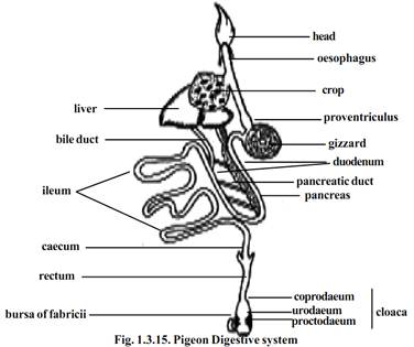
The alimentary canal proper starts from the Pharynx. The Pharynx leads into a long oesophagus that runs back through the neck. At the base of the neck region, it enlarges into a thin walled, distensible sac known as crop containing mucous glands. It serves as a store house for the food. The crop is followed by the stomach. The stomach is divisible into two parts, the anterior tubular proventriculus containing gastric glands and a posterior laterally compressed gizzard. The gizzard has a thick muscular wall and a horny inner lining. Its cavity is small and contains small stones which are helpful to grind the food. Thus the gizzard acts as a grinding mill. This type of arrangement is necessary because of the absence of teeth in the buccal cavity. The intestine arises from the right side of the gizzard. It is divisible into an anterior U- shaped duodenum, and a posterior long coiled ileum. The ileum enlarges posteriorly into a short rectum or large intestine. Anteriorly, the rectum bears a pair of small rectal caeca. The rectum opens to the exterior by the cloaca.
Internally the cloaca is divided into three chambers, the anterior coprodaeum, the middle urodaeum and the posterior proctodaeum. The rectum opens into the coprodaem. The urinogenital ducts open into the urodaem. The proctodaem opens to the exterior by a transverse slit like aperture called cloaca. At the proctodacum, there is a dorsal glandular sac known as Bursa of Fabricii. Its function is unknown.

The digestive glands associated with the alimentary canal are the liver and the pancreas. The liver is bilobed with a large right and a small left lobe. It is devoid of gall bladder. There are two bile ducts. They are forms one from each lobe. They open into the duodenum independently. The pancreas lies between the two limbs of the duodenum. It has three ducts, all opening into the distal limb of the duodenum.
Respiratory System:-
The flight activity requires a continuous and abundant supply of oxygen. Hence, the respiratory system of pigeon is highly developed and well differentiated. The respiratory system consists of external nostrils, glottis, larynx, trachea, bronchus and lungs.
The external nostrils are a pair of slit like apertures occurring at the base of upper beak. They communicate to the pharynx by internal nostrils. A glottis lies behind the tongue. It opens into the larynx. The larynx opens into a trachea. The trachea is a long, cylindrical and flexible tube running back- ward through the neck. On entering the thoracic cavity, the trachea expands into a syrinx or voice box. Later it divides into two bronchi, one for each lung. The walls of tracheal and bronchial tubes are supported by a series of closely set cartilagenous rings. Each bronchus enters a bright red lung. The bronchus divides and subdivides into smaller branches, ultimately ending in fine air capillaries. Lungs are solid spongy organs. They do not hang freely in the thoracic cavity but are lodged firmly in the ribs. Some of the bronchial tubes pass through the lungs and communicate with the air cavities in the bone. There are nine air sacs. They are a median interclavicular, a pair of cervical, two pairs of thoracic and a pair of abdominal air sacs.

The air sacs help to maintain high body temperatures. They make the body lighter and help in flight.
Mechanism of Respiration:-
In birds the expiration is an active process. The process of inspiration is passive. In a resting bird, the sternum is moved up and down with the help of intercostal and the abdominal muscles.
During flight, the sternum is rendered immovable due to the support of wings, but the body cavity is raised and lowered by the action of wings and by the lowering of the vertebral column.
Circulatory system:-
The heart is four chambered, with two auricles and two ventricles. There is complete separation of the oxygenated and non-oxygenated blood. Birds have two distinct circulations as arterial and venous systems.

From the right ventricle arises the pulmonary artery carrying deoxygenated blood to the lungs for purification. An aorta arises from the left ventricle (right systemic). It carries oxygenated blood to various regions of the body through several arteries.
Venous system:-
The deoxygenated blood from various regions of the body is collected by several veins. Finally these veins take the blood to the right auricle through the two precaval and a single postcaval veins.

Nervous system:
The brain is divisible into the fore-, mid- and hind brains. The cerebral hemispheres are distinct. They are round and large in size. The olfactory lobes are very small and they do not contain cavities. The diencephalon is hidden from the view by the forward prolongation of the cerebellum. The diencephalon has the pineal body dorsally and infundibulum and pituitary body ventrally. The optic lobes are lateral in position owing to the large size of the cerebral hemispheres and cerebellum. The medulla oblongata instead of being continued backwards as in other tetrapods, descends almost vertically from the cerebellum

Sense organ :-
In pigeon the olfactory sense is poor. There is no external ear. The tympanum is slightly sunken from the surface of the skin.

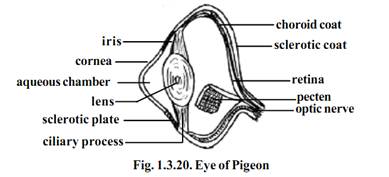
The eyes are large. During flight the eyes and their shape are protected by unique sclerotic plates of the outer eye layer. The nictitating membrane slides over the eyeball and presumably protects the cornea by closing it, during flight. Inside the eye, a vascular pigmented process projects into the vitreous body. It is known as the pecten. It arises from the point of entry of the optic nerve into the eye ball. Its function is not definitely known, but possibly it may help in long distance vision.
Urinogenital system:-
The excretory organs are a pair of kidneys. They are dark red, three lobed structures. They open separately into the urodaeum of the cloaca through two different ureters. There is no urinary bladder. The urine is excreted in the form of uric acid, a semi solid white mass discharged along with faeces through the cloacal aperture.

Male Reproductive system:
The male has a pair of oval testes. From each testis, a duct, the vasdeferens, passes back and opens into the cloaca. The vas deferens is dilated at its posterior end into a seminal vesicle. There is no copulatory organ.
Female reproductive organs:
Only the left ovary persists in the adult. The right ovary disappears during development. The ovary and the oviduct of only one side are functional during breeding season.
References
T. Sargunam Stephen, Biology (Zoology). First Edition – 2005, © Government of Tamilnadu.


|
|
|
|
دراسة يابانية لتقليل مخاطر أمراض المواليد منخفضي الوزن
|
|
|
|
|
|
|
اكتشاف أكبر مرجان في العالم قبالة سواحل جزر سليمان
|
|
|
|
|
|
|
المجمع العلمي ينظّم ندوة حوارية حول مفهوم العولمة الرقمية في بابل
|
|
|