

 النبات
 الحيوان
 الأحياء المجهرية
 علم الأمراض
 التقانة الإحيائية
 التقنية الحيوية المكروبية
 التقنية الحياتية النانوية
 علم الأجنة
 الأحياء الجزيئي
 علم وظائف الأعضاء
 الغدد
الغدد
 المضادات الحيوية
|
Read More
Date: 2-3-2016
Date: 21-3-2016
Date: 2-3-2016
|
Vibrio, Aeromonas, and Plesiomonas
Vibrio cholerae is the most important species in this group from a medical point of view. Cholera vibrios are Gram-negative, comma-shaped, monotrichously flagellated rods. They show alkali tolerance (pH 9), which is useful for selective culturing of V. cholerae in alkaline peptone water. The primary cholera pathogen is serovar O:1. NonO:1 strains (e.g., O:139) cause the typical clinical picture in rare cases. O:1 vibrios are further subdivided into the bio-vars cholerae and eltor. The disease develops when the pathogens enter the intestinal tract with food or drinking water in large numbers (-108). The vi¬brios multiply in the proximal small intestine and produce an enterotoxin. This toxin stimulates a series of reactions in enterocytes, the end result of which is increased transport of electrolytes out of the enterocytes, whereby water is also lost passively. Massive watery diarrhea (up to 20 l/day) results in exsiccosis. The initial therapeutic focus is thus on replacement of lost electrolytes and water. Cholera occurs only in humans. Preventive measures concentrate on protection from exposure to the organism. A killed whole cell vaccine and an attenuated live vaccine are available. They provide only a moderate degree of protection over a period of only six months. International healthcare sources report an incubation period of five days.
The bacteria in these groups are Gram-negative rods with a comma or spiral shape. Their natural habitat is in most cases damp biotopes including the ocean. Some of them cause infections in fish (e.g., Aeromonas salmonicida). By far the most important species in terms of human medicine is Vibrio cholerae.
Vibrio cholerae (Cholera)
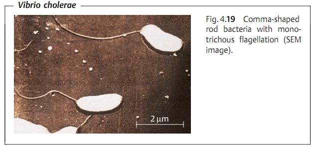
Morphology and culture. Cholera vibrios are Gram-negative rod bacteria, usually slightly bent (comma-shaped), 1.5-2 µm in length, and 0.3-0.5 µm wide, with monotrichous flagellation (Fig. 4.19).
Culturing of V. cholerae is possible on simple nutrient mediums at 37 °C in a normal atmosphere. Owing to its pronounced alkali stability, V. cholerae can be selectively cultured out of bacterial mixtures at pH 9.
Antigens and classification. V. cholerae bacteria are subdivided into serovars based on their O antigens (lipopolysaccharide antigens). The serovar pathogen is usually serovar O:1. Strains that do not react to an O:1 antiserum are grouped together as nonO:1 vibrios. NonO:1 strains were recently described in India (O:139) as also causing the classic clinical picture of cholera. O:1 vibrios are further subclassified in the biovars cholerae and eltor based on physiological characteristics. The var eltor has a very low level of virulence.
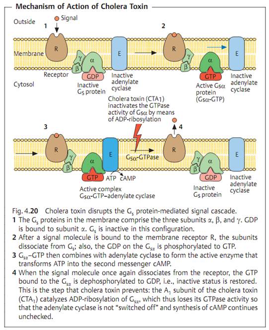
Cholera toxin. Cholera toxin is the sole cause of the clinical disease. This sub-stance induces the enterocytes to increase secretion of electrolytes, above all Cl- ions, whereby passive water loss also occurs. The toxin belongs to the group of AB toxins (see p. 16). Subunit B of the toxin binds to enterocyte receptors, the active toxin subunit A causes the adenylate cyclase in the enterocytes to produce cAMP continuously and in large amounts (Fig. 4.20). cAMP in turn acts as a second messenger to activate protein kinase A, which then activates the specific cell proteins that control secretion of electrolytes. The toxin genes ctxA and ctxB are components of the so-called CTX element, which is integrated in the nucleoid of toxic cholera vibrios (see lysogenic conversion, p. 186) as part of the genome of the filamentous prophage CTXf. The CTX element also includes several regulator genes that regulate both production of the toxin and formation of the so-called toxin-coregulated pili (TCP) on the surface of the Vibrio cells.


Pathogenesis and clinical picture. Infection results from oral ingestion of the pathogen. The infective dose must be large (-108), since many vibrios are killed by the hydrochloric acid in gastric juice. Based on their pronounced stability in alkaline environments, vibrios are able to colonize the mucosa
of the proximal small intestine with the help of TCP (see above) and secrete cholera toxin (see Fig. 4.20). The pathogen does not invade the mucosa.
The incubation period of cholera is two to five days. The clinical picture is characterized by voluminous, watery diarrhea and vomiting. The amount of fluids lost per day can be as high as 20 l. Further symptoms derive from the resulting exsiccosis: hypotension, tachycardia, anuria, and hypothermia. Lethality can be as high as 50% in untreated cases.
Diagnosis requires identification of the pathogen in stool or vomit. Sometimes a rapid microscopical diagnosis succeeds in finding numerous Gram-negative, bent rods in swarm patterns. Culturing is done on liquid or solid selective mediums, e.g., alkaline peptone water or taurocholate gelatin agar. Suspected colonies are identified by biochemical means or by detection of the O:1 antigen in an agglutination reaction.
Therapy. The most important measure is restoration of the disturbed water and electrolyte balance in the body. Secondly, tetracycline and cotrimoxazole can be used, above all to reduce fecal elimination levels and shorten the period of pathogen secretion.
Epidemiology and prevention. Nineteenth-century Europe experienced several cholera pandemics, all of which were caused by the classic cholerae bio- var. An increasing number of cases caused by the biovar eltor, which is characterized by a lower level of virulence, have been observed since 1961. With the exception of minor epidemics in Italy and Spain, Europe, and the USA have been spared major outbreaks of cholera in more recent times. South America has for a number of years been the venue of epidemics of the disease.
Humans are the only source of infection. Infected persons in particular eliminate large numbers of pathogens. Convalescents may also shed V. cholerae for weeks or even months after the infection has abated. Chronic carriers as with typhoid fever are very rare. Transmission of the disease is usually via foods, and in particular drinking water. This explains why cholera can readily spread to epidemic proportions in countries with poor hygiene standards.
Protection from exposure to the pathogen is the main thrust of the relevant preventive measures. In general, control of cholera means ensuring adequate food and water hygiene and proper elimination of sewage. In case of an outbreak, infected persons must be isolated. Infectious excreta and contaminated objects must be disinfected. Even suspected cases of cholera must be reported to health authorities without delay. The incubation period of the cholera vibrio is reported in international health regulations to be five days. A vaccine containing killed cells as well an attenuated live vaccine are available. The level of immunization protection is, however, incomplete and lasts for only six months.
Other Vibrio Bacteria
Vibrio parahemolyticus is a halophilic (salt-friendly) species found in warm ocean shallows and brackish water. These bacteria can cause gastroenteritis epidemics. The pathogen is transmitted to humans with food (seafood, raw fish). The illness is transient in most cases and symptomatic therapy is sufficient.
Vibrio vulnificus is another aquatic organism that produces a very small number of septic infections, mainly in immunosuppressed patients.
Aeromonas and Plesiomonas
The bacteria of these two genera live in freshwater biotopes. Some are capable of causing infection in fish (A. salmonicida). They are occasionally observed as contaminants of moist parts of medical apparatus such as dialysis equipment, vaporizers, and respirators. They can cause nosocomial infections in hospitalized patients with weakened immune systems. Cases of gastroenteritis may result from eating foods contaminated with large numbers of these bacteria.


|
|
|
|
5 علامات تحذيرية قد تدل على "مشكل خطير" في الكبد
|
|
|
|
|
|
|
تستخدم لأول مرة... مستشفى الإمام زين العابدين (ع) التابع للعتبة الحسينية يعتمد تقنيات حديثة في تثبيت الكسور المعقدة
|
|
|