

 النبات
 الحيوان
 الأحياء المجهرية
 علم الأمراض
 التقانة الإحيائية
 التقنية الحيوية المكروبية
 التقنية الحياتية النانوية
 علم الأجنة
 الأحياء الجزيئي
 علم وظائف الأعضاء
 الغدد
الغدد
 المضادات الحيوية
|
Read More
Date: 28-2-2016
Date: 26-2-2016
Date: 25-2-2016
|
Morphology of acute inflammation
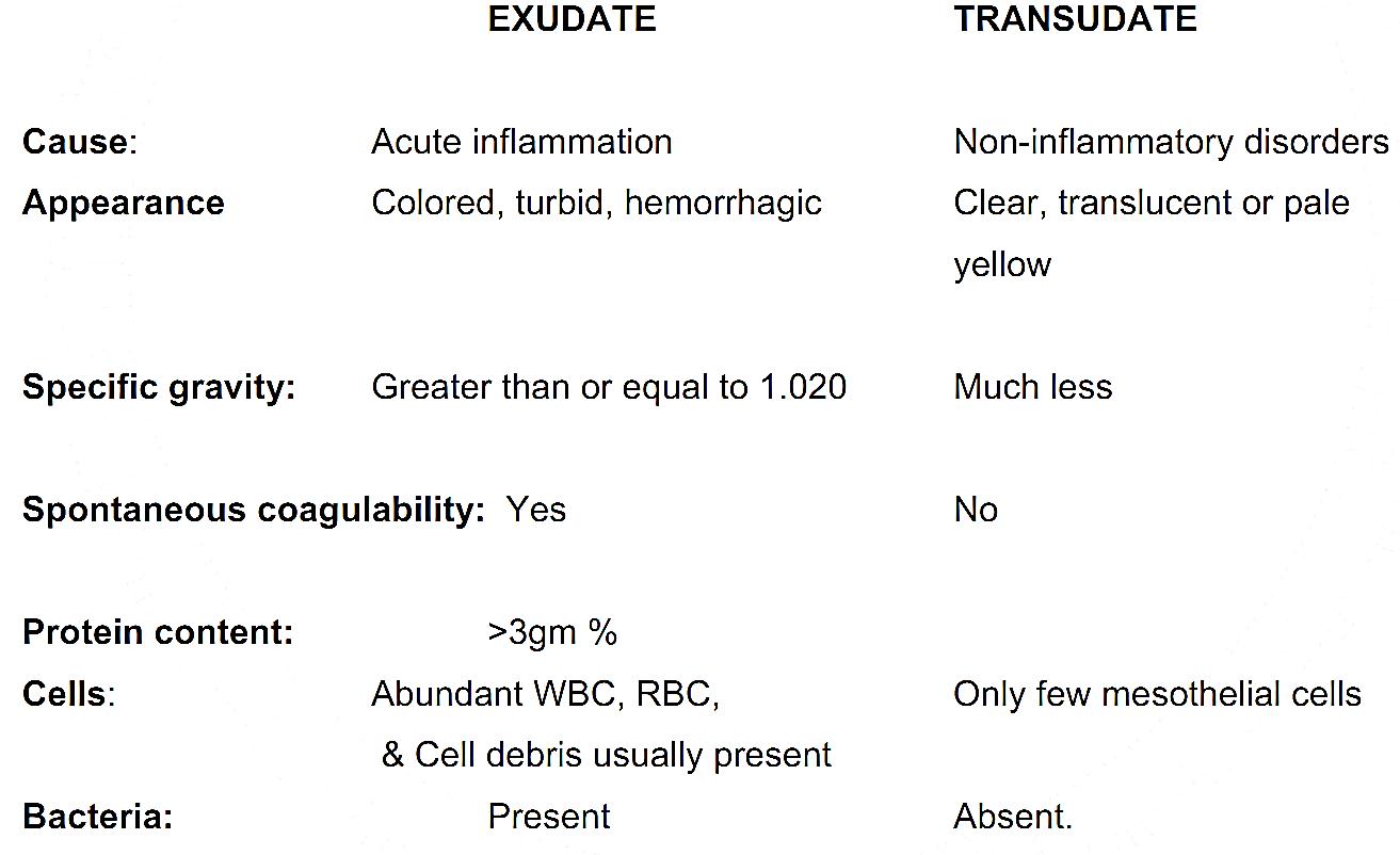
- Characteristically, the acute inflammatory response involves production of exudates. An exudate is an edema fluid with high protein concentration, which frequently contains inflammatory cells.
- A transudate is simply a non-inflammatory edema caused by cardiac, renal, undernutritional, & other disorders.
The differences between an exudate and a transudate are

There are different morphologic types of acute inflammation:
1) Serous inflammation
- This is characterized by an outpouring of a thin fluid that is derived from either the blood serum or secretion of mesothelial cells lining the peritoneal, pleural, and pericardial cavities.
- It resolves without reactions
2) Fibrinous inflammation
- More severe injuries result in greater vascular permeability that ultimately leads to exudation of larger molecules such as fibrinogens through the vascular barrier.
- Fibrinous exudate is characteristic of inflammation in serous body cavities such as the pericardium (butter and bread appearance) and pleura.
Course of fibrinous inflammation include:
- Resolution by fibrinolysis
- Scar formation between perietal and visceral surfaces i.e. the exudates get organized
- Fibrous strand formation that bridges the pericardial space.
3) Suppurative (Purulent) inflammation
This type of inflammation is characterized by the production of a large amount of pus.
Pus is a thick creamy liquid, yellowish or blood stained in colour and composed of
- A large number of living or dead leukocytes (pus cells)
- Necrotic tissue debris
- Living and dead bacteria
- Edema fluid
There are two types of suppurative inflammation:
A) Abscess formation:
- An abscess is a circumscribed accumulation of pus in a living tissue. It is encapsulated by a so-called pyogenic membrane, which consists of layers of fibrin, inflammatory cells and granulation tissue.
B) Acute diffuse (phlegmonous) inflammation
- This is characterized by diffuse spread of the exudate through tissue spaces. It is caused by virulent bacteria (eg. streptococci) without either localization or marked pus formation. Example: Cellulitis (in palmar spaces).
4) Catarrhal inflammation
- This is a mild and superficial inflammation of the mucous membrane. It is commonly seen in the upper respiratory tract following viral infections where mucous secreting glands are present in large numbers, eg. Rhinitis.
5) Pseudomembranous inflammation
- The basic elements of pseudomembranous inflammation are extensive confluent necrosis of the surface epithelium of an inflamed mucosa and severe acute inflammation of the underlying tissues. The fibrinogens in the inflamed tissue coagulate within the necrotic epithelium. And the fibrinogen, the necrotic epithelium, the neutrophilic polymorphs, red blood cells, bacteria and tissue debris form a false (pseudo) membrane which forms a white or colored layer over the surface of inflamed mucosa.
- Pseudomembranous inflammation is exemplified by Dipthetric infection of the pharynx or larynx and Clostridium difficille infection in the large bowel following certain antibiotic use.
References
Bezabeh ,M. ; Tesfaye,A.; Ergicho, B.; Erke, M.; Mengistu, S. and Bedane,A.; Desta, A.(2004). General Pathology. Jimma University, Gondar University Haramaya University, Dedub University.


|
|
|
|
مخاطر خفية لمكون شائع في مشروبات الطاقة والمكملات الغذائية
|
|
|
|
|
|
|
"آبل" تشغّل نظامها الجديد للذكاء الاصطناعي على أجهزتها
|
|
|
|
|
|
|
المجمع العلميّ يُواصل عقد جلسات تعليميّة في فنون الإقراء لطلبة العلوم الدينيّة في النجف الأشرف
|
|
|