

 النبات
 الحيوان
 الأحياء المجهرية
 علم الأمراض
 التقانة الإحيائية
 التقنية الحيوية المكروبية
 التقنية الحياتية النانوية
 علم الأجنة
 الأحياء الجزيئي
 علم وظائف الأعضاء
 الغدد
الغدد
 المضادات الحيوية
|
Read More
Date: 15-1-2017
Date: 1-8-2016
Date: 3-1-2017
|
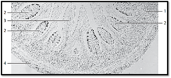
Gut- Associated Lymphoid T issue (GALT)-Dome Areas
Dome areas 2 bulge from the lymphatic tissue 1 of the submucosal tissue into the intestinal lumen. In comparison with the epithelium from mucous membranes of the large intestines 3 , the epithelium over the dome areas shows special attributes: there are no microvilli , crypts or mucous-producing goblet cells. Instead of microvilli, these specialize d epithelial cells show microfolds at their surfaces (microfolds cells, M-cells). M-cells can take up antigenic materials from the lumen and transport it to the lymphatic tissue. M-cells cannot be identified using histological routine preparations and light microscopy. M-cells from a rabbit contain a lot of vimentin as a component of intermediary filaments. It was therefore use d here as a marker for the presence of M-cells. The immunohistochemical localization of vimentin pro-duce d black precipitates in the epithelium. Vermiform appendix of a rabbit.
1 Lymph follicles
2 Dome areas
3 Mucous membrane from the large intestine
4 Tunica muscularis
Stain: immunohistochemical localization of vimentin; differential interference contrast im-aging; magnification: × 80

References
Kuehnel, W.(2003). Color Atlas of Cytology, Histology, and Microscopic Anatomy. 4th edition . Institute of Anatomy Universitätzu Luebeck Luebeck, Germany . Thieme Stuttgart · New York .


|
|
|
|
للعاملين في الليل.. حيلة صحية تجنبكم خطر هذا النوع من العمل
|
|
|
|
|
|
|
"ناسا" تحتفي برائد الفضاء السوفياتي يوري غاغارين
|
|
|
|
|
|
|
نحو شراكة وطنية متكاملة.. الأمين العام للعتبة الحسينية يبحث مع وكيل وزارة الخارجية آفاق التعاون المؤسسي
|
|
|