

 النبات
 الحيوان
 الأحياء المجهرية
 علم الأمراض
 التقانة الإحيائية
 التقنية الحيوية المكروبية
 التقنية الحياتية النانوية
 علم الأجنة
 الأحياء الجزيئي
 علم وظائف الأعضاء
 الغدد
الغدد
 المضادات الحيوية
|
Read More
Date: 25-7-2016
Date: 12-1-2017
Date: 17-1-2017
|
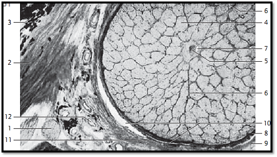
Optic Nerve
Section of the optic nerve behind the lamina cribrosa sclerae.
1 Ciliary nerve
2 Sclera
3 Pigment epithelium of the lamina suprachoroidea
4 Fiber bundles of the optic nerve
5 Central retinal artery
6 Sept a of the piamater
7 Central retinal vein
8 Internal sheath of the optic nerve (piamater)
9 External sheath of the optic nerve (dura mater)
10 Subdural space
11 Arachnoidea
12 Short posterior ciliary artery
Stain: hematoxylin-eosin; magnification: × 20

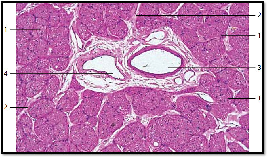
Optic Nerve
Cross-section of the optic nerve behind the lamina cribrosa sclerae. The axons of the ganglia cells are combine d in bundles 1 . They are envelope d by a thin septum of the piamater 2 . The optic nerve contains a piamater sheath, an arachnoidea sheath and a dura mater sheath. The piamater sheath attaches directly to the optic nerve. Septa originate with the piamater and guide blood vessels to the myelinate nerve f ibers. The nerve fiber bundles contain astrocytes and oligodendrocytes. Arteries 3 and central retinal veins 4 are visible in the center of the section. The vessels are sheathed by the loose connective tissue of the piamater.
1 Nerve fiber bundles
2 Piamater septa
3 Central retinal arter y
4 Central retinal vein
Stain: hematoxylin-eosin; magnification: × 40

References
Kuehnel, W.(2003). Color Atlas of Cytology, Histology, and Microscopic Anatomy. 4th edition . Institute of Anatomy Universitätzu Luebeck Luebeck, Germany . Thieme Stuttgart · New York .


|
|
|
|
التوتر والسرطان.. علماء يحذرون من "صلة خطيرة"
|
|
|
|
|
|
|
مرآة السيارة: مدى دقة عكسها للصورة الصحيحة
|
|
|
|
|
|
|
نحو شراكة وطنية متكاملة.. الأمين العام للعتبة الحسينية يبحث مع وكيل وزارة الخارجية آفاق التعاون المؤسسي
|
|
|