

 النبات
 الحيوان
 الأحياء المجهرية
 علم الأمراض
 التقانة الإحيائية
 التقنية الحيوية المكروبية
 التقنية الحياتية النانوية
 علم الأجنة
 الأحياء الجزيئي
 علم وظائف الأعضاء
 الغدد
الغدد
 المضادات الحيوية
|
Read More
Date: 27-7-2016
Date: 9-1-2017
Date: 11-1-2017
|
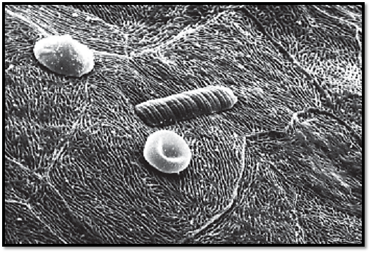
Microvilli-Microplicae
The cell surface can also be enlarged by raised, leaf-like formations of the plasmalemma—i.e., by outward extension in the form of folds. The figure shows flat epithelial cells from the canine tongue with a dense pattern of microplicae —i.e., raise d folds of the plasma membrane. Such microplicae folds are commonly observe d in a multitude of patterns. Microplicae also exist at the bottom face of flat epithelial cells, and therefore may play a role in intercellular adhesion. The more pronounced “edges” in this figure correspond to the cell borders. In transmission electron microscopy, vertical cuts show microplicae of ten as short, stump-shape d microvilli. Located on top of the epithelial cells are two erythrocytes and a rod-shaped form, which can be categorize d as an oral cavity saprophyte.
Scanning electron microscopy; magnification: × 3600

References
Kuehnel, W.(2003). Color Atlas of Cytology, Histology, and Microscopic Anatomy. 4th edition . Institute of Anatomy Universitätzu Luebeck Luebeck, Germany . Thieme Stuttgar t · New York .


|
|
|
|
"عادة ليلية" قد تكون المفتاح للوقاية من الخرف
|
|
|
|
|
|
|
ممتص الصدمات: طريقة عمله وأهميته وأبرز علامات تلفه
|
|
|
|
|
|
|
المجمع العلمي للقرآن الكريم يقيم جلسة حوارية لطلبة جامعة الكوفة
|
|
|