

 النبات
 الحيوان
 الأحياء المجهرية
 علم الأمراض
 التقانة الإحيائية
 التقنية الحيوية المكروبية
 التقنية الحياتية النانوية
 علم الأجنة
 الأحياء الجزيئي
 علم وظائف الأعضاء
 الغدد
الغدد
 المضادات الحيوية
|
Read More
Date: 5-1-2017
Date: 1-8-2016
Date: 14-8-2016
|
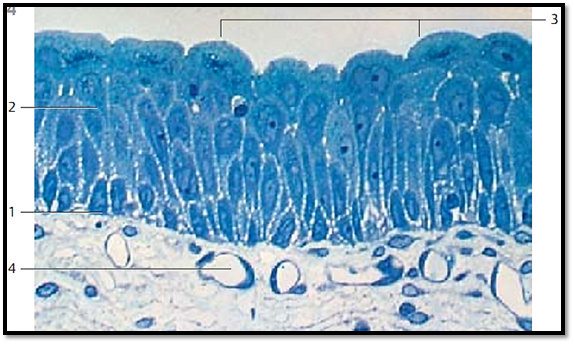
Transitional Epithelium-Urothelium-Ureter
This figure illustrates the basis for the controversy about the configuration of the urothelium. Seemingly, all cells are anchored on the basal membrane. Since they are of different heights, their nuclei appear at different heights from the basal membrane—“multilayered Pseudostratified epithelium.” There are basal 1 , intermediary 2 and superficial cells 3 . Large cells in the superficial layer of ten have two nuclei. They bulge into the lumen, each one covering several intermediary cells—superficial cells. By definition, the superficial cells must be anchored at the basal layer, if seen as a multilayered Pseudostratified epithelium .
1 Basal cells
2 Intermediary cells
3 Cells of the superficial layer
4 Capillaries of the lamina propria
Semi-thin section; stain: methylene blue; magnification: × 350

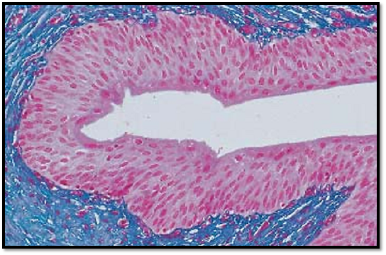
Cross-section through a moderately distended ureter (detail magnification). The transitional epithelium appears as multilayered squamous epithelium. It contains no glands. The superficial cells are flattened . As the bladder expands, the cells of the deeper layers are also flattened. Now, the microfolds at the cell surfaces between epithelial cell junctions are less prominent. Underneath the epithelium, the lamina propria extends as a thick layer of connective tissue with a tight-meshed network of capillaries.
Stain: azan; magnification: × 200

References
Kuehnel, W.(2003). Color Atlas of Cytology, Histology, and Microscopic Anatomy. 4th edition . Institute of Anatomy Universitätzu Luebeck Luebeck, Germany . Thieme Stuttgar t · New York .


|
|
|
|
التوتر والسرطان.. علماء يحذرون من "صلة خطيرة"
|
|
|
|
|
|
|
مرآة السيارة: مدى دقة عكسها للصورة الصحيحة
|
|
|
|
|
|
|
نحو شراكة وطنية متكاملة.. الأمين العام للعتبة الحسينية يبحث مع وكيل وزارة الخارجية آفاق التعاون المؤسسي
|
|
|