

 النبات
 الحيوان
 الأحياء المجهرية
 علم الأمراض
 التقانة الإحيائية
 التقنية الحيوية المكروبية
 التقنية الحياتية النانوية
 علم الأجنة
 الأحياء الجزيئي
 علم وظائف الأعضاء
 الغدد
الغدد
 المضادات الحيوية
|
Read More
Date: 1-12-2015
Date: 15-4-2021
Date: 16-3-2021
|
Colchicine
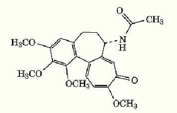
Colchicine (Fig. 1), a compound obtained from the autumn crocus, Colchicum autumnale, is an ancient drug that has been used for centuries for the treatment of gout (1). Its use in molecular biology began in the mid-1930s when its potent ability to inhibit eukaryotic cell proliferation at mitosis was discovered. Since then, colchicine has had a remarkable history as an experimental tool for characterizing the biochemical properties of tubulin, the protein subunit of microtubules, for characterizing the diverse processes in eukaryotic cells that are dependent upon drug-sensitive microtubules, and for studying the polymerization and dynamics properties of microtubules. For example, colchicine has been used to determine the role of microtubules in mitosis, protein secretion, axonal transport, and the development and maintenance of asymmetric cell shape. It has also been used extensively as a cytogenetic tool to determine chromosome numbers in karyotypes. Also, radiolabeled colchicine was used as an affinity marker to effect the first purification of tubulin from brain (2).

Figure 1. Structure of colchicine.
1. Colchicine Binding to Tubulin
The binding of colchicine to tubulin is not a simple process (3-6). The binding reaction is slow and highly temperature-dependent. Binding is extremely slow at 0°C, and 2 to 3 h is required to reach equilibrium at 37°C. The activation energy for the forward reaction is high, ~20 kcal/mol. The kinetics of binding are biphasic. The initial step is the formation of a reversible preequilibrium complex that is followed by a slow step in which conformational changes in tubulin lead to the formation of a final-state tubulin-colchicine (TC) complex. Although the binding of colchicine to tubulin is noncovalent, the final state TC complex is very poorly reversible.
Colchicine binds to tubulin at a single site with a dissociation constant in the range of 0.02 µM to 5 µM. Colchicine has very different affinities for tubulin isolated from different sources. For example, colchicine binds strongly to vertebrate brain tubulin, but it binds very weakly to plant, fungal, and yeast tubulins. Also, there are different tubulin isotypes, and colchicine binds differently to the isotypes purified from same tubulin source (7). Despite intensive investigation, the precise location in tubulin of the colchicine-binding site is not clear. The binding site is thought to reside in the b-tubulin subunit, near residues Cys 354 and Cys 241, close to the intradimer interface.
The binding of colchicine to tubulin induces conformational changes in tubulin, as well as in colchicine (6, 8, 9). For example, the binding of colchicine to tubulin quenches the intrinsic tryptophan fluorescence of tubulin, indicating that it induces a small change in the tertiary structure of the protein. It also perturbs the far-ultraviolet circular dichroism spectrum of tubulin, indicating that it changes the secondary structure of the protein. In addition, colchicine binding to tubulin strongly increases the intrinsic GTPase activity of tubulin, increases the affinity of the ab dimer association by threefold, changes the exposure of thiol groups in tubulin, and, under certain conditions, induces tubulin to self-assemble into nonmicrotubule polymeric structures. Tubulin is also subject to a time- and temperature-dependent irreversible decay of its protein structure. Binding of colchicine to tubulin slows the rate of decay. The concept that colchicine undergoes conformational changes upon binding to tubulin is evident from the development of colchicine-tubulin fluorescence and the change of the colchicine circular dichroism spectrum upon binding to the protein.
2. Inhibition of Microtubule Polymerization by Colchicine
Colchicine inhibits microtubule polymerization at concentrations that are far below the total concentration of tubulin (10, 11), indicating that colchicine inhibits microtubule polymerization by acting at the microtubule ends. In order to produce its potent actions on microtubule polymerization, colchicine must first form a TC complex. Substoichiometric concentrations of TC-complex only partially depolymerize microtubules, and relatively low concentrations of TC-complex can stabilize microtubules against dilution-induced disassembly (12). These studies support the hypothesis that the TC-complex forms a stabilizing “cap” at the end of the microtubule. Also, the TC-complex can form copolymers with unliganded tubulin when microtubules are assembled in the presence of TC complex (13).
3. Kinetic Suppression of Microtubule Dynamics
Microtubules exhibit two kinds of nonequilibrium dynamics: treadmilling and dynamic instability. Recent studies have revealed that colchicine and other compounds that depolymerize microtubules strongly suppress these dynamics at relatively low concentrations in the absence of appreciable microtubule depolymerization. Colchicine was found some years ago to suppress treadmilling in vitro and the rate of microtubule disassembly upon dilution of the microtubules [now called “kinetic capping” (11, 12)]. However, its stabilizing effects on dynamics were only fully appreciated with the introduction of real-time differential-interference contrast video microscopy, which enabled one to visualize directly in real time the stabilizing action of the drug on the growing and shortening dynamics of individual microtubules (14). Small numbers of incorporated TC complexes strongly suppress the rates and extents of growing and shortening and greatly increase the percentage of time that the microtubules spend in an attenuated state. In addition, the TC complex strongly suppresses the catastrophe frequency and increases the rescue frequency. At low submicromolar concentrations, TC complex suppresses the dynamics without reducing the polymer mass. Significant reduction of polymer mass requires relatively high TC complex concentrations. However, the surviving microtubules are extremely stable. Colchicine appears to suppress microtubule dynamics by binding at the microtubule ends, most probably by inducing a conformational change and/or by steric hindrance at the ends.
References
1. P. Dustin (1984). Microtubels, Springer-Verlag, Berlin, pp. 1–482.
2. R. C. Weisenberg, G. G. Borisy, and E. W. Taylor (1968) Biochemistry 7, 4466–4478.
3. G. G. Borisy and E. W. Taylor (1967) J. Cell. Biol. 34, 525–533.
4. L. Wilson and M. Friedkin (1967) Biochemistry 6, 3126–3135.
5. B. Bhattacharyya and J. Wolff (1974) Proc. Natl. Acad. Sci. USA 71, 2627–2631.
6. D. L. Garland (1978) Biochemistry 17, 4266–4272.
7. R. F. Luduena (1983) Mol. Biol. Cell 4, 445–457.
8. J. M. Andreu and S. N. Timasheff (1982) Biochemistry 21, 6465–6476.
9.T. David-Pfeuty, C. Simon, and D. Pantaloni (1979) J. Biol. Chem. 254, 11696–11702.
10. J. B. Olmsted and G. G. Borisy (1973) Biochemistry 12, 4282–4289.
11.R. L. Margolis and L. Wilson (1977) Proc. Natl. Acad. Sci. USA 74, 3466–3478.
12. R. L. Margolis, C. T. Rauch, and L. Wilson (1980) Biochemistry 19, 5550–5557.
13.H. Sternlicht and I. Ringel (1979) J. Biol. Chem. 254, 10540–10550.
14. D. Panda, J. E. Daijo, M. A. Jordan, and L. Wilson (1995) Biochemistry 34, 9921–9929.


|
|
|
|
"عادة ليلية" قد تكون المفتاح للوقاية من الخرف
|
|
|
|
|
|
|
ممتص الصدمات: طريقة عمله وأهميته وأبرز علامات تلفه
|
|
|
|
|
|
|
المجمع العلمي للقرآن الكريم يقيم جلسة حوارية لطلبة جامعة الكوفة
|
|
|