آخر المواضيع المضافة

 النبات
الحيوان
الأحياء المجهرية
علم الأمراض
التقانة الإحيائية
التقنية الحيوية المكروبية
التقنية الحياتية النانوية
علم الأجنة
الأحياء الجزيئي
علم وظائف الأعضاء
الغدد
المضادات الحيوية
النبات
الحيوان
الأحياء المجهرية
علم الأمراض
التقانة الإحيائية
التقنية الحيوية المكروبية
التقنية الحياتية النانوية
علم الأجنة
الأحياء الجزيئي
علم وظائف الأعضاء
الغدد
المضادات الحيوية| Differentiation and Functions of CD8+ Cytotoxic T Lymphocytes |


|
|
Read More
Date: 2025-01-19
Date: 2025-02-02
Date: 2025-03-25
|
Phagocytes are best at killing microbes that are confined to vesicles, and microbes that directly enter the cytosol (e.g., viruses) or escape from phagosomes into the cytosol (e.g., some ingested bacteria) are relatively resistant to the microbicidal mechanisms of phagocytes. Eradication of such cytosolic pathogens requires another effector mechanism of T cell–mediated immunity: CD8+ CTLs. CTLs also serve a vital role in defense against cancers .
CD8+ T lymphocytes activated by antigen and other signals differentiate into CTLs that are able to kill infected cells expressing the antigen. Naive CD8+ T cells can recognize antigens but are not capable of killing antigen-expressing cells. The differentiation of naive CD8+ T cells into fully active CTLs is accompanied by the synthesis of molecules involved in cell killing, giving these effector T cells the functional capacity that is the basis for their designation as cytotoxic. CD8+ T lymphocytes recognize class I MHC–associated peptides on infected cells and tumor cells. The sources of class I– associated peptides are protein antigens synthesized in the cytosol and protein antigens of phagocytosed microbes that escape from phagocytic vesicles into the cytosol . In addition, some dendritic cells may capture the antigens of infected cells and tumors, transfer these antigens into the cytosol, and thus present the ingested antigens on class I MHC molecules, by the process known as cross-presentation (Fig. 1). The differentiation of naive CD8+ T cells into functional CTLs and memory cells requires not only anti gen recognition but also costimulation and, in some situations, help from CD4+ T cells ( Fig. 2).

Fig1. Class I MHC-restricted cross-presentation of microbial antigens from infected cells by dendritic cells. Fragments of cells infected with intracellular microbes (e.g., viruses) or antigens produced in these cells are ingested by dendritic cells, and the antigens of the infectious microbes are broken down and presented in association with class I MHC molecules of the antigen-presenting cells (APCs). T cells recognize the microbial antigens expressed on the APCs, and the T cells are activated. By convention, the term cross-presentation (or cross-priming) is applied to CD8+ T cells (cytotoxic T lymphocytes) recognizing class I MHC–associated antigens (as shown); the same cross-presenting APC may display class II MHC–associated antigens from the microbe for recognition by CD4+ helper T cells.

Fig2. Proteins of the B7 and CD28 families. Ligands on APCs that are homologous to B7 bind to receptors on T cells that are homologous to CD28. Different ligand-receptor pairs serve distinct roles in immune responses. CD28 and ICOS are stimulatory receptors on T cells, and CTLA-4 and PD-1 are inhibitory receptors. Their functions are discussed in the text.
CD8+ CTLs recognize class I MHC–peptide complexes on the surface of infected cells and kill these cells, thus eliminating the reservoir of infection. The T cells recognize MHC-associated peptides by their TCR and the CD8 coreceptor. These infected cells also are called targets of CTLs, because they are destroyed by the CTLs. The TCR and CD8, as well as other signaling proteins, cluster in the CTL membrane at the site of contact with the target cell and are surrounded by the leukocyte function–associated antigen 1 (LFA-1) integrin. These molecules bind their ligands on the target cell, forming an immune synapse .
Antigen recognition by CTLs results in the activation of signal transduction pathways that lead to the exocytosis of the contents of the CTL’s granules into the synapse between the CTL and the target cell (Fig. 3). Because all nucleated cells express class I MHC, and differentiated CTLs do not require costimulation or T cell help for activation, the CTLs can be activated by and are able to kill any infected cell in any tissue. CTLs kill target cells mainly as a result of delivery of granule proteins into the target cells. Two types of granule proteins critical for killing are granzymes (granule enzymes) and perforin. Perforin disrupts the integrity of the target cell plasma membrane and endosomal membranes, thereby facilitating the delivery of granzymes into the cytosol. Granzymes (granule enzymes) cleave and thereby activate enzymes called caspases (cysteine proteases that cleave proteins after aspartic acid residues) that are present in the cytosol of target cells and whose major function is to induce apoptosis.

Fig3. Mechanisms of killing of infected cells by CD8+ cytotoxic T lymphocytes (CTLs). CTLs recognize class I major histocompatibility complex (MHC)–associated peptides of cytoplasmic microbes in infected cells and form tight adhesions (conjugates) with these cells. Adhesion molecules such as integrins stabilize the binding of the CTLs to infected cells (not shown). The CTLs are activated to release (exocytose) their granule contents (perforin and granzymes) toward the infected cell, referred to as the target cell. Granzymes are delivered to the cytosol of the target cell by a perforin-dependent mechanism. Granzymes then induce apoptosis. ICAM-1, Intercellular adhesion molecule 1; LFA-1, leukocyte function–associated antigen 1.
Activated CTLs also express a membrane protein called Fas ligand, which binds to a death-inducing receptor, called Fas (CD95), on target cells. Engagement of Fas activates caspases and induces target cell apoptosis; this pathway does not require granule exocytosis and probably plays only a minor role in killing by CD8+ CTLs.
The net result of these effector mechanisms of CTLs is that the infected cells are killed. Cells that have under gone apoptosis are rapidly phagocytosed and eliminated. CTLs themselves are not injured during the process of killing other cells, so each CTL can kill a target cell, detach, and go on to kill additional targets.
In addition to their cytotoxic activity, CD8+ effector cells secrete IFN-γ. This cytokine is responsible for activation of macrophages in infections and in disease states where excessive activation of CD8+ T cells may be a feature. It may also play a role in defense against some tumors.
Although we have described the effector functions of CD4+ T cells and CD8+ T cells separately, these types of T lymphocytes may function cooperatively to destroy intracellular microbes (Fig. 4). If microbes are phagocytosed and remain sequestered in macrophage vesicles, CD4+ T cells may be adequate to eradicate these infections by secreting IFN-γ and activating the microbicidal mechanisms of the macrophages. However, if the microbes are able to escape from vesicles into the cytoplasm, they become insusceptible to the killing mechanisms of activated macrophages, and their elimination requires destruction of the infected cells by CD8+ CTLs.

Fig4. Cooperation between CD4+ and CD8+ T cells in eradication of intracellular infections. In a macrophage infected by an intracellular bacterium, some of the bacteria are sequestered in vesicles (phagosomes), and others may escape into the cytosol. CD4+ T cells recognize antigens derived from the vesicular microbes and activate the macrophage to kill the microbes in the vesicles. CD8+ T cells recognize antigens derived from the cytosolic bacteria and are needed to kill the infected cell, thus eliminating the reservoir of infection. CTL, Cytotoxic T lymphocyte; IFN, interferon.
|
|
|
|
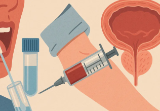
التوتر والسرطان.. علماء يحذرون من "صلة خطيرة"
|
|
|
|
|
|
|
مرآة السيارة: مدى دقة عكسها للصورة الصحيحة
|
|
|
|
|
|
|
نحو شراكة وطنية متكاملة.. الأمين العام للعتبة الحسينية يبحث مع وكيل وزارة الخارجية آفاق التعاون المؤسسي
|
|
|