


النبات

مواضيع عامة في علم النبات
الجذور - السيقان - الأوراق
النباتات الوعائية واللاوعائية
البذور (مغطاة البذور - عاريات البذور)
الطحالب
النباتات الطبية
الحيوان
مواضيع عامة في علم الحيوان
علم التشريح
التنوع الإحيائي
البايلوجيا الخلوية
الأحياء المجهرية
البكتيريا
الفطريات
الطفيليات
الفايروسات
علم الأمراض
الاورام
الامراض الوراثية
الامراض المناعية
الامراض المدارية
اضطرابات الدورة الدموية
مواضيع عامة في علم الامراض
الحشرات
التقانة الإحيائية
مواضيع عامة في التقانة الإحيائية
التقنية الحيوية المكروبية
التقنية الحيوية والميكروبات
الفعاليات الحيوية
وراثة الاحياء المجهرية
تصنيف الاحياء المجهرية
الاحياء المجهرية في الطبيعة
أيض الاجهاد
التقنية الحيوية والبيئة
التقنية الحيوية والطب
التقنية الحيوية والزراعة
التقنية الحيوية والصناعة
التقنية الحيوية والطاقة
البحار والطحالب الصغيرة
عزل البروتين
هندسة الجينات
التقنية الحياتية النانوية
مفاهيم التقنية الحيوية النانوية
التراكيب النانوية والمجاهر المستخدمة في رؤيتها
تصنيع وتخليق المواد النانوية
تطبيقات التقنية النانوية والحيوية النانوية
الرقائق والمتحسسات الحيوية
المصفوفات المجهرية وحاسوب الدنا
اللقاحات
البيئة والتلوث
علم الأجنة
اعضاء التكاثر وتشكل الاعراس
الاخصاب
التشطر
العصيبة وتشكل الجسيدات
تشكل اللواحق الجنينية
تكون المعيدة وظهور الطبقات الجنينية
مقدمة لعلم الاجنة
الأحياء الجزيئي
مواضيع عامة في الاحياء الجزيئي
علم وظائف الأعضاء
الغدد
مواضيع عامة في الغدد
الغدد الصم و هرموناتها
الجسم تحت السريري
الغدة النخامية
الغدة الكظرية
الغدة التناسلية
الغدة الدرقية والجار الدرقية
الغدة البنكرياسية
الغدة الصنوبرية
مواضيع عامة في علم وظائف الاعضاء
الخلية الحيوانية
الجهاز العصبي
أعضاء الحس
الجهاز العضلي
السوائل الجسمية
الجهاز الدوري والليمف
الجهاز التنفسي
الجهاز الهضمي
الجهاز البولي
المضادات الميكروبية
مواضيع عامة في المضادات الميكروبية
مضادات البكتيريا
مضادات الفطريات
مضادات الطفيليات
مضادات الفايروسات
علم الخلية
الوراثة
الأحياء العامة
المناعة
التحليلات المرضية
الكيمياء الحيوية
مواضيع متنوعة أخرى
الانزيمات

Ulcerative colitis
 المؤلف:
James Carton
المؤلف:
James Carton
 المصدر:
Oxford Handbook of Clinical Pathology 2024
المصدر:
Oxford Handbook of Clinical Pathology 2024
 الجزء والصفحة:
3rd edition , p134-135
الجزء والصفحة:
3rd edition , p134-135
 2025-02-15
 1191
1191


+
-
20
Definition
• An idiopathic inflammatory bowel disease, characterized by inflammation restricted to the large bowel mucosa, which always involves the rectum and extends proximally in a continuous fashion for a variable distance.
Epidemiology
• Uncommon.
• Major incidence between 15 and 25 y.
Aetiology and pathogenesis
• thought to be due to an abnormal mucosal immune response to luminal bacteria.
• the genetic link is weaker than for Crohn’s disease (CD).
• Smoking appears to decrease the risk of UC.
• One unusual, but consistently confirmed, observation is the protective effect of appendectomy on the subsequent development of UC.
Presentation
• recurrent episodes of bloody diarrhoea, often with urgency and tenesmus.
Macroscopy
• erythematous mucosa with a friable, eroded surface and haemorrhage.
• Inflamed mucosa may form polypoid projections (inflammatory polyps).
• Disease always involves the rectum and extends continuously to involve a variable amount of colon (fig. 1).
Histopathology
• Biopsies show almost always show evidence of chronic mucosal damage: crypt architectural distortion, Paneth cell metaplasia, and loss of the inflammatory cell gradient in the lamina propria. there is also mucosal inflammation with cryptitis and crypt abscess formation. Inflammation. the changes are usually more marked distally although the rectal changes may be mild if there has been topical treatment.
• resection specimens show diffuse inflammation limited to the mucosal layer. Inflammatory polyps may be present.
• extension of inflammation into the submucosa or muscle layers may occur in very severe acute UC, but the inflammation still remains heaviest in the mucosal layer.
Prognosis
• Generally good with treatment.
• Increased risk of colorectal carcinoma, so surveillance colonoscopy is usually recommended several years after diagnosis.
• extra- GI manifestations include enteropathic arthropathy , primary sclerosing cholangitis , erythema nodosum , pyoderma gangrenosum , uveitis, and AA amyloidosis.
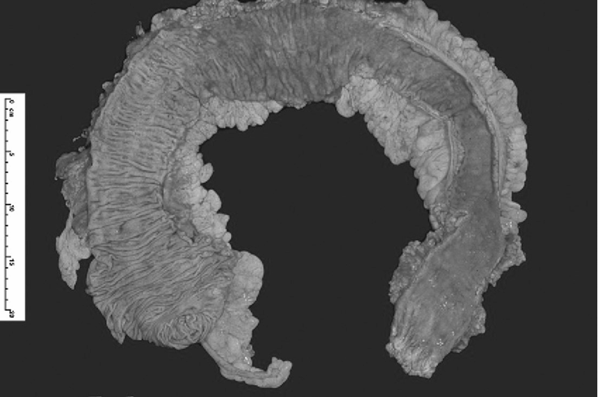
fig1. Ulcerative colitis. this is a colectomy specimen from a patient with ulcerative colitis. the right colon is on the left of the picture (note the appendix), and the left colon and rectum are on the right side of the picture. the inflamed mucosa, which looks red, begins at the rectum and continuously affects the left colon until the transverse colon where there is a sharp transition into normal mucosa . reproduced with permission from Clinical Pathology (Oxford Core texts), Carton, James, Daly, richard, and ramani, Pramila, Oxford University Press (2006), p. 163, figure 8.15.
 الاكثر قراءة في مواضيع عامة في علم الامراض
 اخر الاخبار
اخبار العتبة العباسية المقدسة

الآخبار الصحية


مواضيع ذات صلة
 قسم الشؤون الفكرية يصدر كتاباً يوثق تاريخ السدانة في العتبة العباسية المقدسة
 "المهمة".. إصدار قصصي يوثّق القصص الفائزة في مسابقة فتوى الدفاع المقدسة للقصة القصيرة
 (نوافذ).. إصدار أدبي يوثق القصص الفائزة في مسابقة الإمام العسكري (عليه السلام)


