
DNA and nanomaterials
 المؤلف:
Peter Atkins, Tina Overton, Jonathan Rourke, Mark Weller, and Fraser Armstrong
المؤلف:
Peter Atkins, Tina Overton, Jonathan Rourke, Mark Weller, and Fraser Armstrong
 المصدر:
Shriver and Atkins Inorganic Chemistry ,5th E
المصدر:
Shriver and Atkins Inorganic Chemistry ,5th E
 الجزء والصفحة:
ص681-682
الجزء والصفحة:
ص681-682
 2025-10-15
 304
304
DNA and nanomaterials
Key point: Interactions between gold nanoparticles and DNA can cause the self-assembly of DNA into condensates and the ordering of nanoparticles into regular arrays that can be used for biosensors. DNA condensates are self-assembled nanostructures. The architecture of these condensates is driven by the components that become woven into the nanoassembly. Electrostatic interactions drive the formation of DNA condensates. DNA is a negatively charged polyelectrolyte that interacts with positively charged ions, molecules, or modified nanoparticles to form ordered structures and convert from their random coils to a more compact form. This compact form, called a condensate, is essential for the organization of DNA into chromatin, a component of genes. Mimicking or controlling the DNA condensation process is the basis of the design of nonviral gene delivery vectors that have practical application in the treatment of cancer, drug delivery, and biosensing. Studies of DNA condensation in vitro have focused on the use of complex cations, including hexaamminecobalt (III) or polyamines as the positive charge centre in the condensate. Complex formation in these systems can be expected to mimic DNA wrapping around charged particles in living cells, such as positively charged histone proteins. Because the cationic particles can be fine-tuned with respect to size and charge density, the effect of those parameters on the complex formation patterns with DNA have begun to be understood. Gold nanoparticles have been suggested as an effective transfection agent (a device for introducing exogeneous DNA into a cell) and, as such, it is important to understand the interactions of nanoparticles with DNA. Figure 25.37 shows AFM images of complexes of lysine-modified gold nanoparticles and DNA at different nanoparticle/DNA ratios. These results indicate the possibility of developing

Figure 25.37 AFM images of complexes of lysine-modified gold nanoparticles and DNA at different nanoparticle/DNA ratios (a) 10:1, (b) 20:1, (c) 50:1, and (d) 100:1. Several wrapped molecules are seen along with free nanoparticles in (a) and (b). Network formation is seen in (c) and (d). The inset in (a) shows a single DNA molecule wrapping around six nanoparticles. (Reproduced by permission from M. Ganguli, et al., Langmuir, 2004, 20, 5165.)
functionalized gold nanoparticle-DNA into a model system to study DNA condensation in vivo. These smart nanomaterials have far-reaching consequence for the design of nanoscale self-assembled materials that can be used as building blocks for active nanodevices. Another exciting accomplishment in the synthesis of bionanomaterials is the use of DNA to drive the assembly of inorganic nanoparticles (Fig. 25.38). First, nanoparticles are functionalized with single strands of DNA. Then, the complementary strand of DNA is introduced from an analyte or bioagent, leading to dimerization with the DNA on the nanoparticles and the self-assembly of the nanoparticles into ordered arrays. Detectable differencesin optical properties occur between the disordered and ordered states and result in colour changes. This work has helped to make sophisticated optical-based biosensor arrays with remarkable sensitivity and selectivity for different analytes and bioagents.
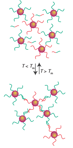
Figure 25.38 DNA templating of inorganic semiconducting nanoparticles. (N.L. Rosi and C.A. Mirkin, Chem. Rev., 2005, 105, 1547.)
 الاكثر قراءة في مواضيع عامة في الكيمياء العضوية
 اخر الاخبار
اخبار العتبة العباسية المقدسة


