


النبات

مواضيع عامة في علم النبات
الجذور - السيقان - الأوراق
النباتات الوعائية واللاوعائية
البذور (مغطاة البذور - عاريات البذور)
الطحالب
النباتات الطبية
الحيوان
مواضيع عامة في علم الحيوان
علم التشريح
التنوع الإحيائي
البايلوجيا الخلوية
الأحياء المجهرية
البكتيريا
الفطريات
الطفيليات
الفايروسات
علم الأمراض
الاورام
الامراض الوراثية
الامراض المناعية
الامراض المدارية
اضطرابات الدورة الدموية
مواضيع عامة في علم الامراض
الحشرات
التقانة الإحيائية
مواضيع عامة في التقانة الإحيائية
التقنية الحيوية المكروبية
التقنية الحيوية والميكروبات
الفعاليات الحيوية
وراثة الاحياء المجهرية
تصنيف الاحياء المجهرية
الاحياء المجهرية في الطبيعة
أيض الاجهاد
التقنية الحيوية والبيئة
التقنية الحيوية والطب
التقنية الحيوية والزراعة
التقنية الحيوية والصناعة
التقنية الحيوية والطاقة
البحار والطحالب الصغيرة
عزل البروتين
هندسة الجينات
التقنية الحياتية النانوية
مفاهيم التقنية الحيوية النانوية
التراكيب النانوية والمجاهر المستخدمة في رؤيتها
تصنيع وتخليق المواد النانوية
تطبيقات التقنية النانوية والحيوية النانوية
الرقائق والمتحسسات الحيوية
المصفوفات المجهرية وحاسوب الدنا
اللقاحات
البيئة والتلوث
علم الأجنة
اعضاء التكاثر وتشكل الاعراس
الاخصاب
التشطر
العصيبة وتشكل الجسيدات
تشكل اللواحق الجنينية
تكون المعيدة وظهور الطبقات الجنينية
مقدمة لعلم الاجنة
الأحياء الجزيئي
مواضيع عامة في الاحياء الجزيئي
علم وظائف الأعضاء
الغدد
مواضيع عامة في الغدد
الغدد الصم و هرموناتها
الجسم تحت السريري
الغدة النخامية
الغدة الكظرية
الغدة التناسلية
الغدة الدرقية والجار الدرقية
الغدة البنكرياسية
الغدة الصنوبرية
مواضيع عامة في علم وظائف الاعضاء
الخلية الحيوانية
الجهاز العصبي
أعضاء الحس
الجهاز العضلي
السوائل الجسمية
الجهاز الدوري والليمف
الجهاز التنفسي
الجهاز الهضمي
الجهاز البولي
المضادات الميكروبية
مواضيع عامة في المضادات الميكروبية
مضادات البكتيريا
مضادات الفطريات
مضادات الطفيليات
مضادات الفايروسات
علم الخلية
الوراثة
الأحياء العامة
المناعة
التحليلات المرضية
الكيمياء الحيوية
مواضيع متنوعة أخرى
الانزيمات

The Sensory Organs
 المؤلف:
T. Sargunam Stephen
المؤلف:
T. Sargunam Stephen
 المصدر:
Biology (Zoology)
المصدر:
Biology (Zoology)
 الجزء والصفحة:
 5-11-2015
 2779
2779


+
-
20
The Sensory Organs
Living organism respond to several stimuli such as light, heat, sound. chemicals, pressure, touch, stretch and orientation. These stimuli are felt by specific receptors. The receptors convert the stimuli into impulses in the nervous systems.
The touch receptors in the skin are the simplest receptors. Such receptors are single nerve cells responding directly to the stimulus. Other receptors are complex sense organs. On these organs the stimulus is channeled into a receptive region of the organ. Among the several organs, the most important are the eyes and ears.
The eye
The eye is formed of 3 coats or tunics.
Coats or tunic Regions
. 1. Outer or fibrous - sclera & cornea
. 2. Middle or vascular - choroid, ciliary body & iris
. 3. Inner or nervous – retina
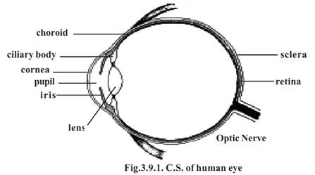
The sclera is the white outer layer of the eye. It covers posterior five- sixths of the eye. This firm layer provides shape and protects the internal structures. A small region of the sclera can be seen as the “white of the eye”.
In the front, the outer layer forms a transparent region called the cornea. It permits entry of light. The cornea is made up of a connective tissue having collagen, elastic fibres and proteoglycans.
The middle tunic of the eyeball is the vascular tunic. It contains most of the blood vessels. The vascular tunic contains melanin containing pigment cells. It appears black in colour. A major part of the vascular tunic is found in association with the sclera and called the choroid. Anteriorly this layer forms the ciliary body and iris.
The ciliary body consists of smooth muscles called the ciliary muscles. Contraction of the ciliary muscles can change the shape of the lens.
The iris is the coloured part of the eye. It may be black, brown or blue. It is a contractile structure surrounding an opening called the pupil. Light enters the eye through the pupil. The iris regulates such entry by controlling the size of the pupil.
The inner most tunic of the eye is the retina. It consists of an outer pigmented retina and an inner sensory retina. The sensory retina is light sensitive. It contains nearly 120 million photoreceptor cells called rods and another 7 million cones.
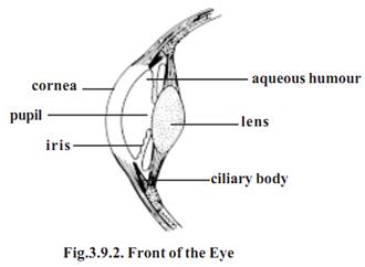
Compartments of the eye: The eye has 2 major compartments. There is a smaller compartment anterior to the lens. Behind the lens there is a larger compartment.
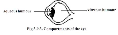
The anterior compartment is divided into two chambers. There is an anterior chamber found between the cornea and iris. A smaller posterior chamber lies between the iris and lens. These two chambers are filled with a substance called the aqueous humor. It helps to maintain intraocular pressure.
The posterior compartment of the eye is much larger and it contains a transparent jellylike substance called vitreous humor.
The eye lens is a unique biological structure. It is transparent and biconvex. It is made up of long columnar epithelial cells called lens fibres. These fibres have an accumulation of proteins called crystalline. The lens is placed between the two eye compartments by suspensory ligaments.
The functioning of the eye is aided by accessory structures. They include the eyebrows, eyelids, conjunctiva and lacrimal apparatus.
The eyebrows prevent the sweat during perspiration from running down into the eye. They help to shade the eyes from direct sunlight.
The eyelids and associated lashes protect the eyes from foreign objects. The medial region where the eyelids join has a small reddish-pink mound called the caruncle. It contains modified sebaceous and sweat glands. There are two or three rows of hairs attached to the free edges of eyelids. Modified sweat glands called the ciliary glands open into the follicles of the eyelashes. It keeps them lubricated. The inner margin of the eyelids contain Melbomian glands. These glands produce sebum for lubricating the eyelids.
The inner surface of the eyelids and the anterior surface of the eye are covered by a thin, transparent mucous membrane called the conjunctiva.
The lacrymal glands or tear glands are situated in the super lateral corner of the eye orbit. They produce tear at the rate of about 1 ml / day. It helps to moisten the eye surface and wash away foreign substances. At the corners of the eye there are small openings called the puncta. Each punctum in turn opens into a lacrymal canaliculus. The lacrimal canaliculi open into a lacrimal sac. This sac enters into a nasolacrimal duct which opens into the inferior nasal concha. These ducts help to drain the excess tear. The entire organization related to ‘tear’ is called the lacrymal apparatus.
Ears (The organs of hearing)
The ears are the organs of hearing and balance. They have three parts, namely external, middle and inner ears.
External ear - The fleshy part outside the head is called the pinna. It is made up of elastic cartilage and skin. It is followed by the external auditory meatus. This passage is lined with hairs and ceruminous glands. These glands produce cerumen or earwax. The hair and wax prevent foreign objects from reaching the ear drum. The ear drum or tympanic membrane is a oval, three layered structure. It separates outer and middle ears.
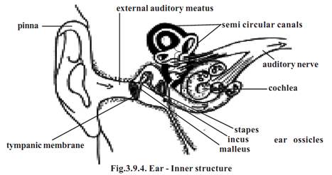
Middle ear - It is an air filled cavity. It contains three auditory ossicles called the malleus, incus and stapes. The handle of malleus is in contact with the inner surface of the ear drum. The head of the malleus is attached to the incus. While the stapes on one side is attached to the incus, its other side fits into the oval window. The oval window leads to the inner ear.
Inner ear - This region has tunnels and chambers inside the temporal bone called the bony labyrinth. The bony labyrinth contains three regions called the cochlea, vestibule and semicircular canals. The oval window found in between the middle and inner ears communicates with the vestibule of the inner ear. The organs of the inner ear perceive the sound.
References
T. Sargunam Stephen, Biology (Zoology). First Edition – 2005, Government of Tamilnadu.
 الاكثر قراءة في علم التشريح
 اخر الاخبار
اخبار العتبة العباسية المقدسة

الآخبار الصحية


 قسم الشؤون الفكرية يصدر كتاباً يوثق تاريخ السدانة في العتبة العباسية المقدسة
 "المهمة".. إصدار قصصي يوثّق القصص الفائزة في مسابقة فتوى الدفاع المقدسة للقصة القصيرة
 (نوافذ).. إصدار أدبي يوثق القصص الفائزة في مسابقة الإمام العسكري (عليه السلام)


