آخر المواضيع المضافة

 النبات
الحيوان
الأحياء المجهرية
علم الأمراض
التقانة الإحيائية
التقنية الحيوية المكروبية
التقنية الحياتية النانوية
علم الأجنة
الأحياء الجزيئي
علم وظائف الأعضاء
الغدد
المضادات الحيوية
النبات
الحيوان
الأحياء المجهرية
علم الأمراض
التقانة الإحيائية
التقنية الحيوية المكروبية
التقنية الحياتية النانوية
علم الأجنة
الأحياء الجزيئي
علم وظائف الأعضاء
الغدد
المضادات الحيوية| Pigments-Purkinje Cells |


|
|
Read More
Date: 2025-04-09
Date: 8-1-2017
Date: 28-12-2020
|
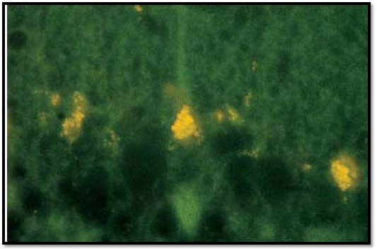
Pigments-Purkinje Cells
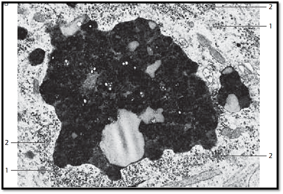
The enzymatic degradation of endocytosed material is often incomplete.
Vesicles and vacuoles with such material are called residual bodies . Lipofuscin is a membrane-enclose d, indigestible residue that is left over after lysosomal degradation. Lipofuscin-loaded residual bodies are called lipofuscin granules. In long-live d muscle and nerve cells, lipofuscin granules b ecome more abundant with age. Lipofuscin is therefore known as an aging or “wear-and-tear” pigment. Lipofuscin granules in Purkinje cells from rat cerebellum. The small lipofuscin granules show an endogenous yellow fluorescence. They appear in small piles at the upper cell pole between nucleus and dendrite. Small granules can occur anywhere in the perikaryon.
Stain: Einarson gallocyanine; fluorescence microscopy (BG-12 excitation filter, 530-nm barrier filter); magnification: × 1000
This figure shows the ultrastructure of a lipofuscin granule. Note the bizarre shape (lipofuscin has an irregular surface), the osmiophilic electron-dense matrix, which consists of numerous very small granules, and the differences in electron densities. In the immediate vicinity of the granule, there are areas with polysomes 1 as well as short ergastoplasmic lamellae 2.
1 Polysomes
2 Nissl bodies
References
Kuehnel, W.(2003). Color Atlas of Cytology, Histology, and Microscopic Anatomy. 4th edition . Institute of Anatomy Universitätzu Luebeck Luebeck, Germany . Thieme Stuttgar t · New York .

|
|
|
|
للعاملين في الليل.. حيلة صحية تجنبكم خطر هذا النوع من العمل
|
|
|
|
|
|
|
"ناسا" تحتفي برائد الفضاء السوفياتي يوري غاغارين
|
|
|
|
|
|
|
بمناسبة مرور 40 يومًا على رحيله الهيأة العليا لإحياء التراث تعقد ندوة ثقافية لاستذكار العلامة المحقق السيد محمد رضا الجلالي
|
|
|