آخر المواضيع المضافة

 النبات
الحيوان
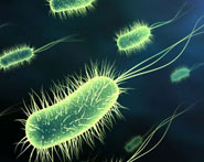
الأحياء المجهرية
علم الأمراض
التقانة الإحيائية
التقنية الحيوية المكروبية
التقنية الحياتية النانوية
علم الأجنة
الأحياء الجزيئي
علم وظائف الأعضاء
الغدد
المضادات الحيوية
النبات
الحيوان
الأحياء المجهرية
علم الأمراض
التقانة الإحيائية
التقنية الحيوية المكروبية
التقنية الحياتية النانوية
علم الأجنة
الأحياء الجزيئي
علم وظائف الأعضاء
الغدد
المضادات الحيوية| Extraepithelial Glands-Mixed (Seromucous) Glands |


|
|
Read More
Date: 1-8-2016
Date: 10-1-2017
Date: 3-8-2016
|
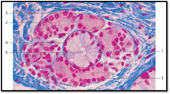
Extraepithelial Glands-Mixed (Seromucous) Glands
This figure displays a mucous terminal portion (tubulus) 1 . It is flanked by several serous acini 2 . The nuclei of serous acinar cells are round 3 while the nuclei of mucous tubule are more flattened 4 . The cytoplasm of serous gland cells is stained red, that of mucous gland cells is light. Note the wide lumen 5 of the mucous tubule.
1 Mucous tubule
2 Serous acini
3 Nuclei of serous gland cells
4 Nuclei of mucous gland cells
5 Lumen of a mucous tubule
Mixed glands from the mucous membrane of the uvula.
Stain: azan; magnification: × 400
References
Kuehnel, W.(2003). Color Atlas of Cytology, Histology, and Microscopic Anatomy. 4th edition . Institute of Anatomy Universitätzu Luebeck Luebeck, Germany . Thieme Stuttgar t · New York .

|
|
|
|
التوتر والسرطان.. علماء يحذرون من "صلة خطيرة"
|
|
|
|
|
|
|
مرآة السيارة: مدى دقة عكسها للصورة الصحيحة
|
|
|
|
|
|
|
نحو شراكة وطنية متكاملة.. الأمين العام للعتبة الحسينية يبحث مع وكيل وزارة الخارجية آفاق التعاون المؤسسي
|
|
|